Progression of coronary artery plaque can eventually narrow the artery enough to significantly limit blood flow to the heart muscle and cause problems. Typical problems include chest pain, difficulty breathing, poor exercise stamina, weak heart performance, and abnormal heart rhythm. Measuring the reduction in coronary artery blood flow is an important way to determine whether opening up a blockage will help the patient.
In November of 2014, the technology company Heartflow (Redwood City, California) earned FDA approval for generating measurements of coronary artery blood flow using specialized analysis of cardiac CT images. The result of this analysis is a series of fractions between 0 and 1 throughout the coronary arteries; these fractions are “fractional flow reserve” values derived from CT (FFR-CT). FFR-CT can tell when a blockage limits coronary artery blood flow and has proven useful to reduce further testing after cardiac CT. High quality cardiac CT images are essential to achieve reliable results from FFR-CT. In May of 2017, we began a working partnership with Heartflow. We are currently the only imaging center in Oklahoma with direct access to FFR-CT for patients referred for cardiac CT.
FFR-CT is most helpful when cardiac CT shows at least “intermediate” coronary artery narrowing. There is generally not a need for FFR-CT when cardiac CT shows no coronary artery disease or mild coronary artery disease.
For an introductory video about FFR-CT and examples of FFR-CT, please see below.
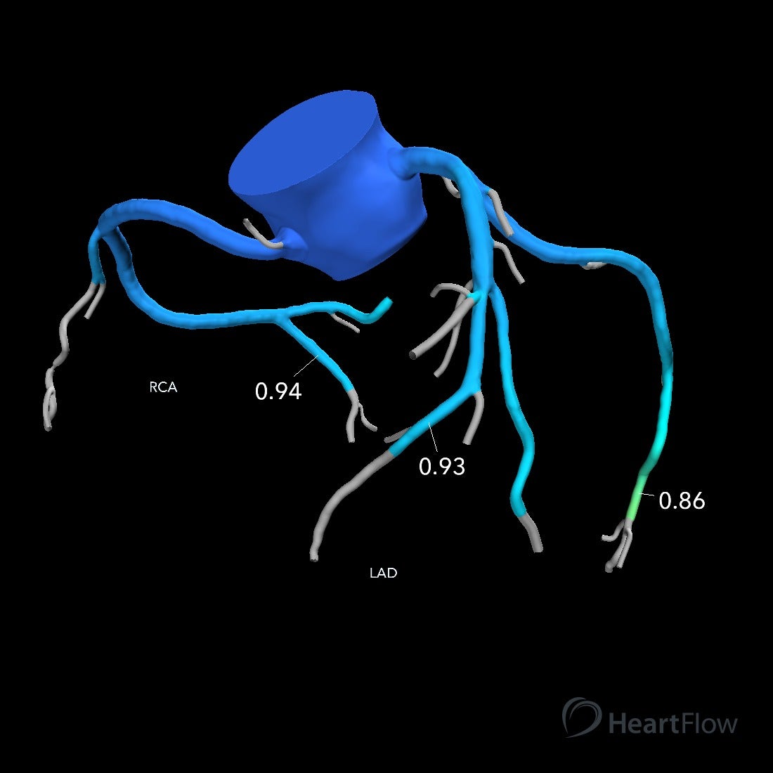
Example of normal coronary artery blood flow by FFR-CT analysis. Note that all values in this example are higher than 0.8; a value below 0.8 is the most commonly accepted criterion for indicating significant blood flow reduction.
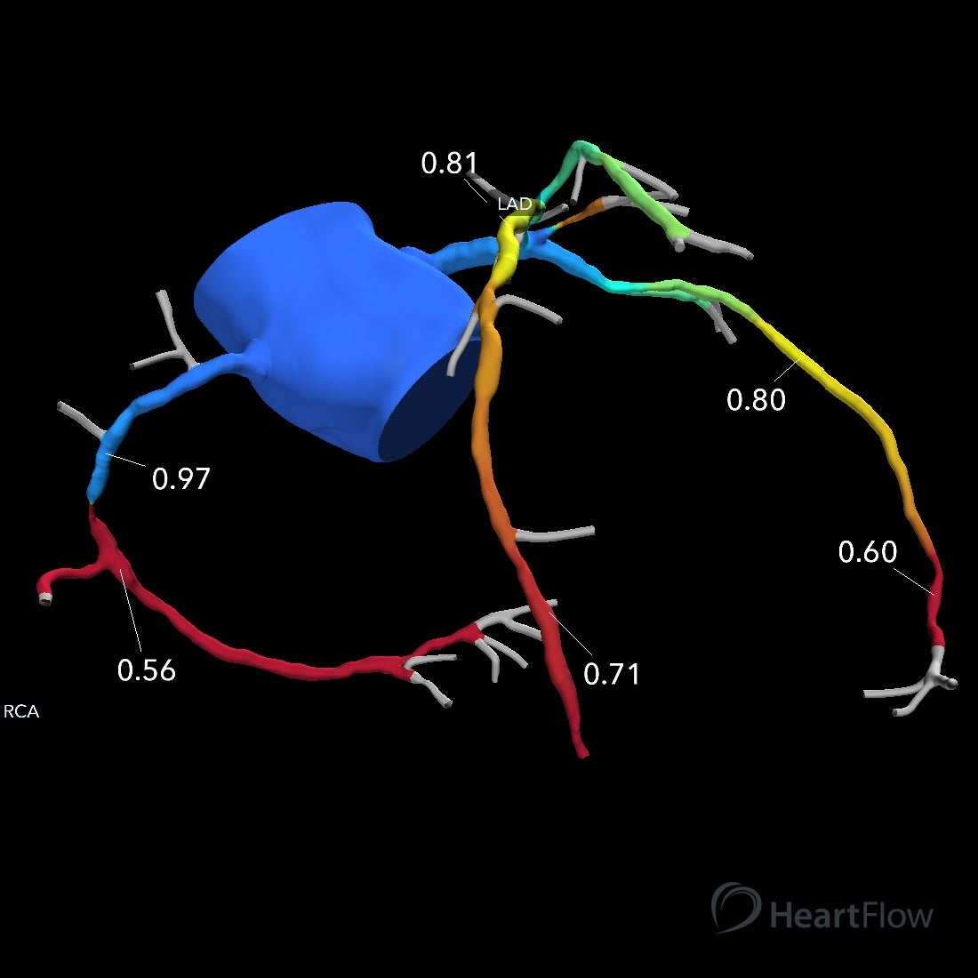
Example of very abnormal coronary artery blood flow by FFR-CT analysis. There are multiple regions with FFR-CT values less than 0.8 (colored as orange or red), demonstrating significant blood flow reduction.